Exploring Green Fluorescent Protein: A Scientific Session with Universiti Sains Malaysia (USM), Malaysia
Jakarta, June 08, 2026 – On May 5th, 2026, i3L University held an online session featuring Dr. Eugene Ong, Ph.D., from Universiti Sains Malaysia, which was moderated by Marchia Rumkorem, i3L International Office. The session introduced students to Green Fluorescent Protein (GFP), covering its discovery, development, and applications in current biological research. It was an interesting opportunity to see how a discovery from nature can become an essential tool in science.
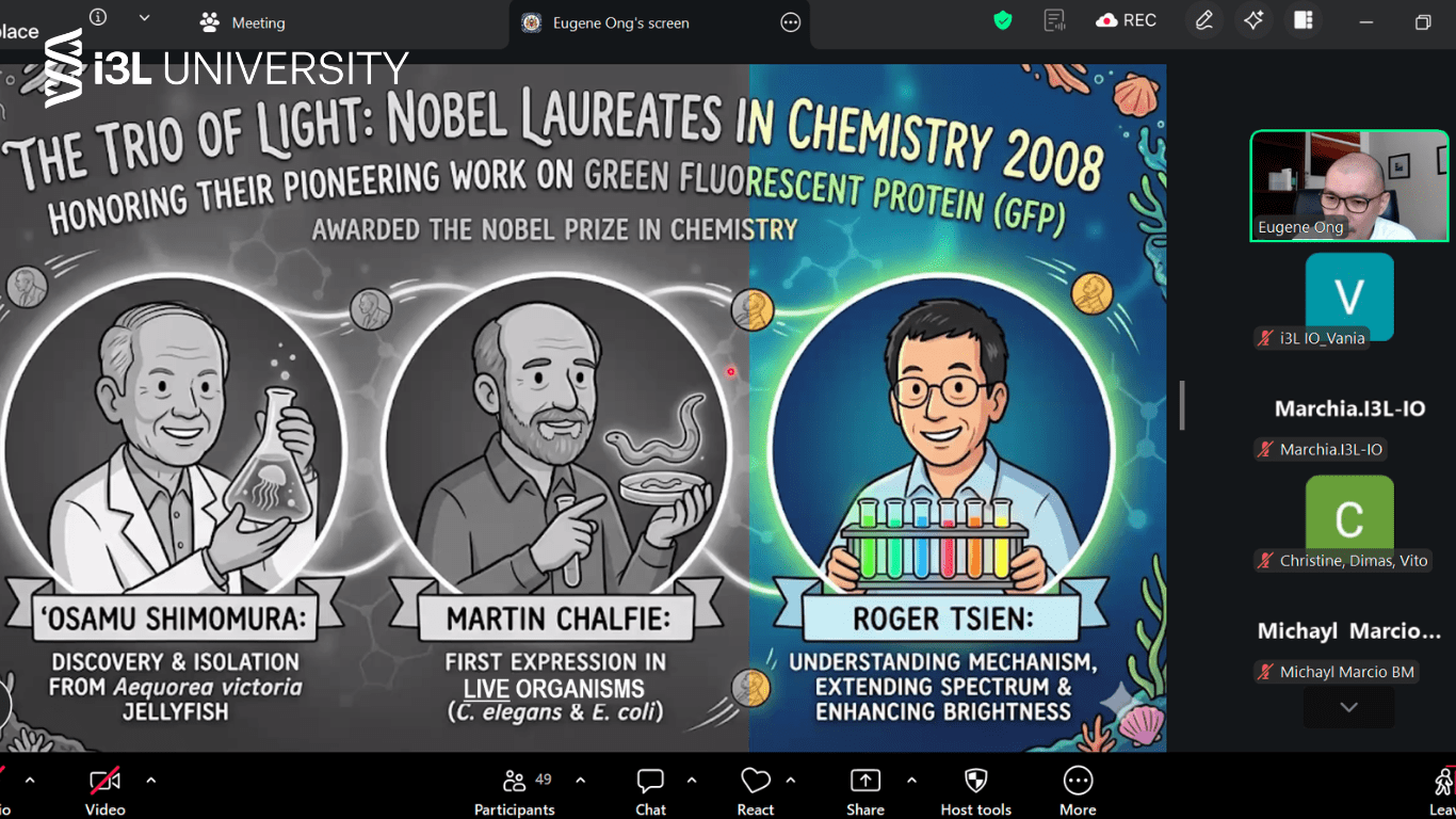
Green Fluorescent Protein (GFP) comes from a deep sea jellyfish called Aequorea victoria. Its discovery led to the 2008 Nobel Prize in Chemistry, awarded to Osamu Shimomura, Martin Chalfie, and Roger Tsien for their important contributions. Osamu Shimomura first identified a glowing protein, which later led to the isolation of aequorin, a protein that emits blue light when it reacts with calcium. GFP then converts this blue light into green fluorescence. Later on, Douglas Prasher successfully cloned the GFP gene in 1992, which made it possible to use GFP in different organisms. After Douglas, Martin Chalfie showed that GFP can form its fluorescent structure on its own, as long as oxygen is present. Meanwhile, Roger Tsien improved GFP through protein engineering, creating different colors and making it brighter and more stable for research use.
The session also discussed how scientists modified fluorescent proteins to improve their performance. Through several modifications, scientists were able to improve its brightness and stability, while also reducing toxicity. These improvements allow researchers to track multiple components in cells at the same time using different colors.
Dr. Ong also explained how GFP is used in his research on bacterial pathogens such as Salmonella Typhi and Leptospira interrogans. These bacteria produce many proteins, and a large number of them still have unknown functions, especially those related to virulence. To study this, researchers use yeast (Saccharomyces cerevisiae) as a model organism. By expressing bacterial proteins in yeast cells, they can observe how these proteins behave and where they are located inside the cell. Different proteins affect the yeast in different ways and localize to different parts of the cell, which helps researchers understand their roles. Moreover, advanced imaging techniques, such as confocal microscopy, are then used to observe these processes more clearly.
Overall, the session showed how GFP has become an important tool in biological research, from its discovery in jellyfish to its use in studying diseases and cellular processes today. Through this session, students were able to see how research develops from basic discoveries into real world applications. Other than that, it also highlights i3L University’s commitment to bringing in global perspectives and up to date scientific knowledge for its students.